
Send us your feedback
Here you can send us feedback on the Maxess-website. Please describe the problem or what’s missing in a clear way, and on what page you found the issue. Thank you so much for your help!
Seeing inside graphene biocomposites with advanced X-ray techniques
Researchers from RISE Research Institutes of Sweden, Biofiber Tech and Holmen used advanced X-ray methods to study how graphene is distributed inside biobased composites. The results provide new tools for quality control and help companies produce more consistent and sustainable materials.
Many companies are developing biobased composites made from renewable raw materials such as wood fibres. By adding graphene, these materials can gain new properties, including better conductivity, strength and barrier performance.
To achieve the desired performance, manufacturers need to control how the graphene is distributed inside the material. In some products, graphene layers should remain separated. In others, a controlled stacking of layers is beneficial. Until now, it has been difficult to distinguish between the structures formed by fibres, polymers and graphene using conventional laboratory methods.
Working Together for Future Solutions
The project brought together Biofiber Tech, Holmen and RISE. Through the BEAMS for Industry programme, the partners gained access to the European Synchrotron Radiation Facility (BM05), (ID13), (ESRF) in France, where powerful X-rays can reveal details that are not visible with standard measurement techniques.
Representatives from the companies took part in both the experiments and the analysis together with researchers and beamline experts.
“The first time we see the 3D visualized bulk structures of biobased fiber composites. Beneficial to product development and demonstration to our customers.”
Luis Valencia, Biofiber Tech AB
A new way to look inside materials
The team combined two complementary techniques. Wide-Angle X-ray Scattering (WAXS) was used to study how graphene layers were arranged at a microscopic level. Micro-computed tomography (µCT) was used to create detailed three-dimensional images of the material without cutting it apart.
Together, these methods provided information about both the internal structure and the distribution of graphene, from tiny features at the microscopic scale to larger structures in finished products.
Key findings
The WAXS measurements showed that graphene formed the desired graphite-like structure in a nanocellulose-based air-barrier film. This result provides an important reference point for future production and quality control.
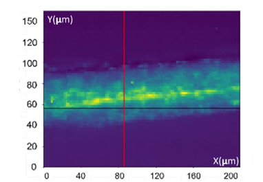
WAXS image that shows bright yellow, revealing that graphene is stacked in a graphite-like air barrier layer.
The analysis also showed that unwanted graphene stacking had been avoided in conductive wood-fibre composites, helping to preserve the material’s functional properties.
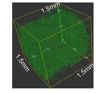
Pore distribution and average fiber size from a 3.4 mm³ µCT volume of 3D-printed WFC kayak.
Using µCT, the researchers created a detailed 3D image of part of a 3D-printed kayak. The data revealed how pores, fibres and material density were distributed throughout the structure, providing valuable information for improving manufacturing processes.
Why this matters
The project shows how advanced X-ray methods can help companies better understand and control complex biobased materials. The results will support the standardisation of air-barrier films, conductive composites and 3D-printed products.
The methods can also be used to study many other sustainable materials where internal structure affects performance. By helping manufacturers see what happens inside their products, these techniques can improve quality, reduce waste and support the development of more resource-efficient manufacturing.
This work was made possible through BEAMS for Industry.
Contact Partners
Case Details
Biofiber Tech, Holmen
RISE offer within large scale research infrastructuresESRF Business Development Office