
Send us your feedback
Here you can send us feedback on the Maxess-website. Please describe the problem or what’s missing in a clear way, and on what page you found the issue. Thank you so much for your help!
Visualizing how coenzyme supplementation aids skin anti-aging
Coenzyme Q10, or Ubiquinone, is an essential cellular component involved in bioenergetic processes. It occurs in many different species besides humans, where it is mostly located in the membranes of cells and mitochondria. Its reduced form, Ubiquinol, has antioxidative properties that help protect the skin, humans’ largest organ, from environmental harms such as UV radiation and pollution. The body’s biosynthesis of Q10 generally decreases with age, resulting in increased cellular oxidative stress. This highlights the importance of understanding Q10 uptake after external administration.
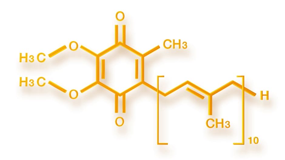
Molecular structure of Coenzyme Q10. (Image: Beiersdorf)
For the first time, scientists from the University of Hamburg have successfully visualized and quantified how cells take up coenzyme Q10 using cutting-edge X-ray fluorescence (XRFUsed for studying the elemental composition of materials with spatial resolution.More info) imaging at DESY’s PETRA III synchrotron. This breakthrough not only reveals how Q10 enters cells but also showcases a powerful, non-invasive tool for studying the distribution of other biomolecules and drugs.
The Challenge: Finding the right method
Coenzyme Q10 cellular uptake after supplementation has remained largely mysterious. Traditional imaging methods like PET or SPECT can track molecules in whole organisms but lack the resolution to see inside individual cells. Other approaches, such as ICP-MS, provide that detail but destroy the sample in the process through its invasive analysis. That’s where imaging at PETRA III comes in — offering both high spatial resolution and non-destructive analysis. This method is non-invasive and by using the facility’s intense, precisely tuned X-rays, the researchers achieved a level of sensitivity that made it possible to quantify Q10 uptake inside living cells.
Illuminating the Invisible: A Synchrotron Breakthrough
To trace Q10’s path, the team created an iodine-labeled version of coenzyme Q10 (I₂-Q10) and introduced it to human skin cells (keratinocytes). After confirming that complementary XRFUsed for studying the elemental composition of materials with spatial resolution.More info experiments:
- At beamline P21.1, whole-cell pellets were scanned, revealing that I₂-Q10 accumulated in specific regions within the individual cells — with an uptake signal roughly three times stronger than that of control samples.
- At beamline P06, sub-cellular mapping was carried out at nanometer resolution. This allowed scientists to pinpoint exactly where Q10 localizes within the cell, switching seamlessly between coarse and fine scans to map hundreds of cells efficiently.
The result: a clear, quantitative picture of Q10’s journey into and through the cell — something no technique had achieved before.
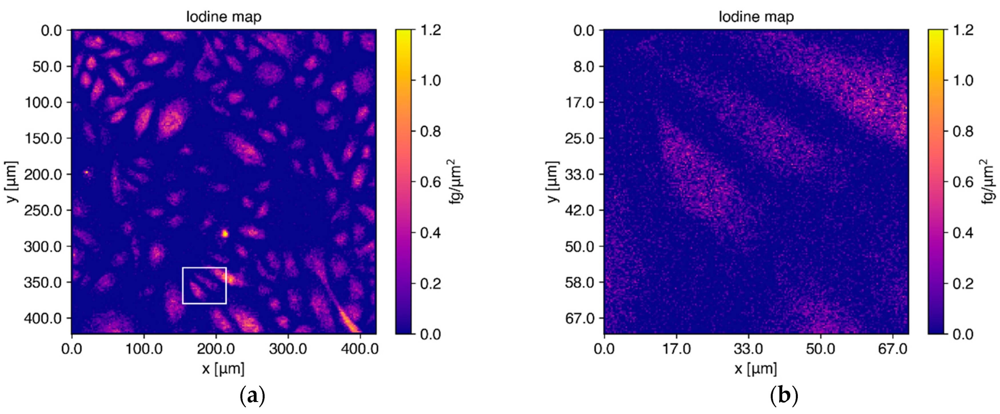
Elemental maps for iodine in cells using XRF at beamline P06. The coarse scan (a) shows how homogeneously distributed iodine is in the I2-Q10 treated cells. The fine scan (b), indicated by the white rectangle in (a), shows a higher resolution and the distribution of the coarse scan in more detail. (Image: DOI 10.3390/antiox11081532)
Enabling biomedical breakthroughs
This study demonstrates that synchrotron-based XRF imaging can precisely track how biomolecules are distributed within cells, without destroying them. The approach paves the way for studying drug delivery, bioavailability, and metabolism at an unprecedented level of detail.
With high photon flux and flexible beamline setups, PETRA III offers a unique platform to explore how molecules behave inside living systems — enabling breakthroughs in both biomedical research and pharmaceutical innovation.
Contact Partners
Case Details
Beiersdorf, DESY
P06 Hard X-ray Micro/Nano-ProbeP21.1 High Energy X-Ray Diffraction – Swedish Beamline